SciencebiologyCellular Biology
“Great Unified Microscope” reveals micro and nano worlds in a single view
KE
Kevin White
2 hours ago7 min read2 comments
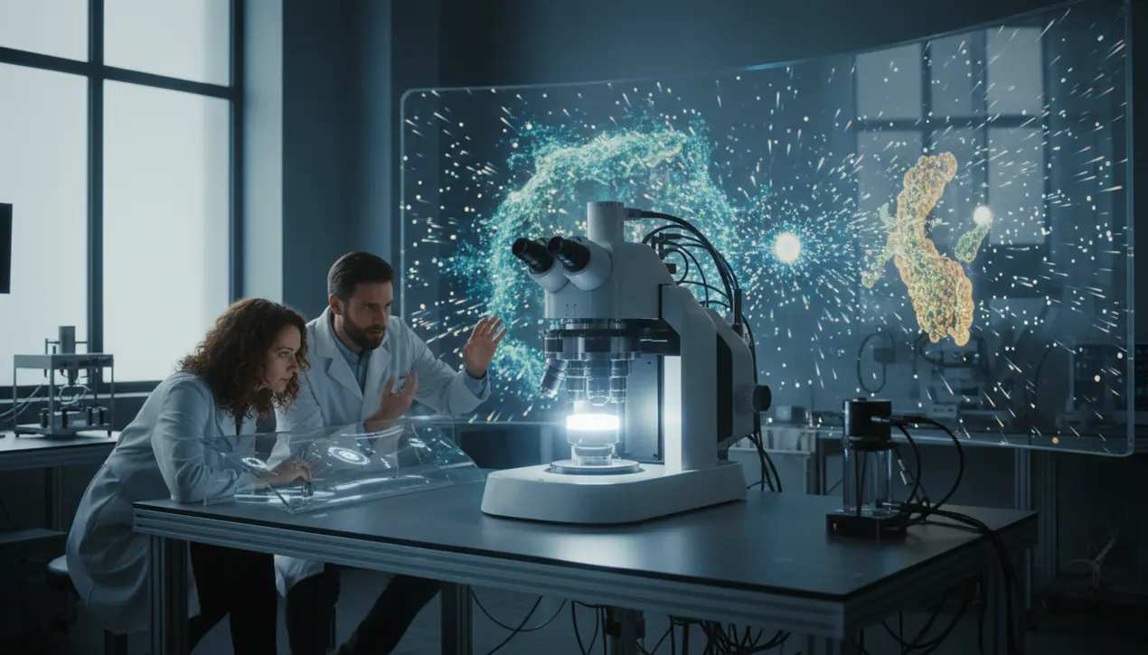
In a breakthrough that feels ripped from the pages of a near-future medical thriller, scientists have unveiled a 'Great Unified Microscope,' a dual-light system that finally bridges the observational chasm between the micro and nano worlds. This isn't just an incremental upgrade; it’s a paradigm shift, allowing researchers to witness the intricate ballet of living cells in real-time without the crude intervention of fluorescent dyes that can perturb the very processes we seek to understand.Imagine watching a city from a helicopter, able to see both the grand sweep of its skyline and the movement of individual cars on its streets—simultaneously. That’s the power of this technology.It captures the detailed architecture of cellular structures while simultaneously tracking the frantic, Brownian dance of minuscule particles, providing a holistic, dynamic view of life at its most fundamental level. The implications are staggering.The creators, in their initial proof-of-concept, turned this new lens on the process of cell death, a critical event in everything from cancer development to neurodegenerative diseases. Traditionally, piecing together this narrative required stitching data from multiple instruments, a bit like trying to understand a film by looking at separate, blurry stills.Now, they can watch the entire feature film in high definition, even estimating the size and refractive index of particles as the cell undergoes its final, programmed shutdown. This quantitative capability moves us from mere observation to precise measurement, a crucial step for diagnostic applications.The next frontier, and the one that truly sets the pulse racing, is the team's ambition to push this technique toward imaging particles as small as viruses. We are standing on the precipice of a new era in virology and immunology.To be able to directly visualize a virus particle—a entity like SARS-CoV-2 that brought the world to a standstill—interacting with a living cell, in its native environment, would be a monumental achievement. It would revolutionize our understanding of infection mechanisms, accelerate antiviral drug discovery by allowing us to see how candidate compounds physically block viral entry, and provide unprecedented insights into the nanoscale machinery of life itself.This development sits at the powerful convergence of AI, advanced optics, and biology—a field sometimes called 'computational microscopy. ' It leverages sophisticated algorithms to interpret the complex light patterns, extracting information that was previously invisible.This is the future of medicine: not just bigger data, but smarter, more integrated visual intelligence. While the current system is a sophisticated research tool, the path to clinical application is clear.One can envision a day when a refined version of this technology in a hospital lab provides rapid, label-free analysis of blood samples, identifying viral loads or characteristic nanoparticles indicative of early-stage cancers long before traditional symptoms appear. The work is a testament to the power of interdisciplinary science, merging physics, engineering, and biology to build a window into a world we have only been able to imagine, until now.
#featured
#microscope
#cellular imaging
#nanotechnology
#cell death
#refractive index
#viruses
Stay Informed. Act Smarter.
Get weekly highlights, major headlines, and expert insights — then put your knowledge to work in our live prediction markets.
Comments
Loading comments...
© 2025 Outpoll Service LTD. All rights reserved.